

Your Order
 FIRST ORDER SPECIAL!
Get 20% OFF on your first order
ORDER NOW
20% OFF FIRST ORDER
FIRST ORDER SPECIAL!
Get 20% OFF on your first order
ORDER NOW
20% OFF FIRST ORDER
Human Anatomy Coursework Help: Reflexes, CNS & Endocrine System
Sample Content
Anatomy and Physiology Coursework: Levers, Muscles, Nervous System, and Endocrine Function
Coursework
Following the American Psychological Association’s Guidelines
Student’s Name
Institutional Affiliation
Date
Referencing Style: APA / Word Count: 1227
Coursework
Answer 1 - Differences between a lever working at a mechanical advantage versus one working at a mechanical disadvantage –
Mechanical Advantage vs. Mechanical Disadvantage of Levers
A lever with a mechanical advantage means that it can overcome or bear large load with little effort (BBC, 2021). Such levers are very useful in joints that have to bear weight of the whole body, such as the Ankle joint.
Mechanical Advantage is expressed as – Effort arm/ Resistance arm (BBC, 2021). It shows that greater the Effort arm in comparison with Resistance arm, the greater the Mechanical advantage. However, when the load arm is longer than the effort arm, it is said to be at a mechanical disadvantage or third-class levers. The example of third-class lever is that of elbow joint (CrossFit, 2019).
Answer 2 - Locations and actions of the muscles typically used in breathing
Locations and Actions of the Respiratory Muscles
There are three groups of respiratory muscles that facilitate respiration in human beings; the diaphragm, the muscles of the rib cage, and abdominal muscles (Aliverti, 2016). These muscles act on the chest wall and its compartments, which are, lung-apposed rib-cage, diaphragm -apposed ribcage and the abdomen respectively (Aliverti, 2016).
The mechanism of breathing involves contraction of the diaphragm, that expands the diaphragm and the lower or abdominal ribcage. Whereas, the rib cage muscles, which are intercostals, parasternals, scalene and the neck muscles act on the upper rib cage part (called pulmonary rib cage) and are both inspiratory as well as expiratory (Aliverti, 2016), the abdominal muscles on the other hand are expiratory as they act on the abdomen and the abdominal ribcage (Aliverti, 2016).
Answer 3 - The Resting Membrane Potential and the impact of K+ on the unequal distribution of charges
Resting Membrane Potential and the Role of K+
The Resting membrane potential is defined as the voltage across the non-signaling (resting) neuron (khanacademy.org, n.d.). This resting potential of a neuron is determined by the permeability of the ions and the concentration gradient across the membrane. Thus, there is a concentration gradient for Na+ and K+ ions also. The movement of ions via channels down the gradient leads to separation of charge, called as Resting Membrane Potential (khanacademy.org, n.d.). Since, the membrane has much more permeability towards K+ than Na+, the resting membrane potential is near to the equilibrium potential of K+. Therefore, the Na+/K+ pump activity and excessive permeability of K+ contributes to the unequal distribution of charges and ions thereby establishing a resting membrane potential of -30 mV to -90 mV (khanacademy.org, n.d.).
Answer 4 - The difference between the temporal and spatial summation in a postsynaptic neuron is as follows (Lakna, 2019):
Temporal vs. Spatial Summation in a Postsynaptic Neuron
| Temporal Summation | Spatial Summation |
| A sensory summation that involves a sum of the stimuli of a number of spatially separated neurons at a time is termed spatial summation |
| Multiple presynaptic neurons are involved in the generation of the action potential. |
| Subthresholds are generated by the multiple presynaptic neurons in this case. |
| It is a more efficient process. |
Answer 5 -
Gross Anatomy of Spinal Cord
The Spinal Cord is the Caudal part of the Central Nervous System. It is protected by the vertebral column, which are the bony structures enclosing the spinal cord. Spinal Cord is covered by the three membranes of the Central Nervous System (CNS), including dura mater, arachnoid and pia mater (Nógrádi and Vrbová, 2013). The spinal cord occupies around two-thirds of the vertebral column in an adult mammal. Retro-caudally, the Spinal Cord is divided into four parts: cervical, thoracic, lumbar and sacral portions. Figure 1 shows gross anatomy of Spinal Cord.
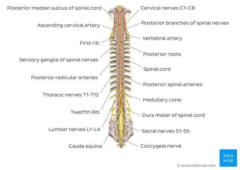
Figure 1
Cross-Sectional Anatomy of Spinal Cord
The cross-sectional view of the Spinal Cord indicates white matter around the butterfly-shaped arrangement of gray matter (Britannica.com, n.d.). The cross-section of the spinal cord shows four surfaces, anterior, posterior and two lateral surfaces (kenhub.com, n.d.). These surfaces have anterior fissures and sulci (posterior, anterolateral, and posterolateral) (kenhub.com, n.d.). the gray matter comprises of neuronal cell bodies, whereas white matter is made of axons (kenhub.com, n.d.). Figure 2 shows cross-sectional image of Spinal cord.
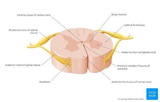
Figure 2
Answer 6 -
Components of Crossed Extensor Reflex
A Crossed Extensor Reflex is a protective withdrawal reflex that helps in protection of body from any harm (unm.edu, n.d.). For instance, when one steps on a nail with the right foot while walking, the reflex action will enable the right leg to withdraw itself from the nail and shift the load on the left leg. The components of reflex involve the extensor muscles (Quadriceps’) relaxation instead of contraction and activation of flexors (hamstrings) to bend the leg and save it from the nail (unm.edu, n.d.), whereas, in the opposite leg muscles, the mechanism will be reversed, that is, contraction of extensors and relaxation of flexors for stabilization of the weight on the left leg. Thus, crossed extensor reflex is a type of withdrawal reflex to protect body from any harm (unm.edu, n.d.).
Answer 7 -
Blood-Brain Barrier Structure and Function
The Blood-Brain Barrier is a barrier between the blood and the Central Nervous System at the level of Brain (Abott et al., 2010). The Blood-Brain Barrier at the micro-vessel endothelium is one of the major-cites of an exchange between blood and CNS. The fenestrated endothelial cells of the barrier facilitate easier passage of water, lipophilic substances and gases across the permeable membrane of the Blood-Brain Barrier. There are also choroidal epithelial cells that produce Cerebrospinal fluid (kenhub.com, 2021).
Answer 8 -
Structural and Functional Relationship Between Hypothalamus and Pituitary Gland
Hypothalamus and the pituitary gland are the main endocrine glands in a human body (Panawala, 2017). Hypothalamus is a small part of the brain that lies beneath the thalamus. The pituitary gland is a gland below the hypothalamus. The hypothalamus is attached with the pituitary gland via the two lobes of the pituitary gland (anterior lobe or adenohypophysis and posterior lobe called neurohypophysis). The hypothalamus connects with the adenohypophysis by a stream of blood whereas connects with the neurohypophysis by means of neurons (Panawala, 2017). The function is thus, based on the structure of the pituitary and hypothalamus, such that the hypothalamus regulates the pituitary gland functions (Panawala, 2017). The hypothalamus-pituitary complex is the ‘command center’ of the entire endocrine system that secrete several hormones like Growth Hormone (GH), Prolactin (PRL), Thyroid-stimulating Hormone (TSH), to name a few (courses.lumenlearning.com, n.d.).
Answer 9 -
Reasons why the sympathetic division of the ANS has more widespread and longer-lasting effects than the parasympathetic division:
The Autonomic Nervous System has two divisions, sympathetic and parasympathetic (courses.lumenlearning.com, n.d.). Whereas, the sympathetic division is responsive for fight-or-flight response, the parasympathetic division on the other hand is known by the epithet of rest and digest (courses.lumenlearning.com, n.d.). Since sympathetic division is more dominant over the parasympathetic division during the times of emotional or physical stress, the effects produced by sympathetic division of ANS are longer lasting and more widespread than the parasympathetic division of the ANS (napavalley.edu, n.d.). Thus, the sympathetic division is overpowering over parasympathetic division during the times of stress and therefore, produce long-lasting and widespread effects.
References
Abbott et al. (2010) Structure and function of the blood-brain barrier. Neurobiol Dis.
Available at https://pubmed.ncbi.nlm.nih.gov/19664713/
Aliverti, A. (2016) The respiratory muscles during exercise. Breathe (Sheff). Available at
https://www.ncbi.nlm.nih.gov/pmc/articles/PMC4933622/
BBC (2021) Movement analysis in sport. Bbc.co.uk. Available at
https://www.bbc.co.uk/bitesize/guides/zwbfg82/revision/2
Courses.lumenlearning.com (n.d.) The Pituitary Gland and Hypothalamus. [online]. Available
at https://courses.lumenlearning.com/suny-ap2/chapter/the-pituitary-gland-and-hypothalamus/
Courses.lumenlearning.com (n.d.) Divisions of the Autonomic Nervous System. [online].
Available at https://courses.lumenlearning.com/ap1/chapter/divisions-of-the-autonomic-nervous-system/
CrossFit (2019) Anatomy of Levers, Part 4: Third-Class Levers. [online]. Available at
https://www.crossfit.com/essentials/anatomy-of-levers-part-4-third-class-levers
Khanacademy.org (n.d.). The membrane potential. [online]. Available at
https://www.khanacademy.org/science/biology/human-biology/neuron-nervous-system/a/the-membrane-potential
Kenhub.com (n.d.) Blood–brain barrier: want to learn more about it? [online]. Available
at https://www.kenhub.com/en/library/anatomy/the-blood-brain-barrier
Kenhub.com (n.d.) Spinal cord. [online]. Available at
https://www.kenhub.com/en/library/anatomy/the-spinal-cord
Lakna (2019) What is the Difference Between Temporal and Spatial Summation. Pediaa.com.
Available at https://pediaa.com/what-is-the-difference-between-temporal-and-spatial-summation/
Napavalley.edu (n.d.) Chapter 20 - Human Anatomy. [online]. Available at
https://www.napavalley.edu/people/briddell/Documents/BIO%20218/Biology_218_Lecture_Outline_20_Autonomic_NS.pdf
Nogradi, A. and Vrbová. G. (2013) Anatomy and Physiology of the Spinal Cord.
Landes Bioscience. Available at https://www.ncbi.nlm.nih.gov/books/NBK6229/
Panawala, L. (2017) Relationship Between Hypothalamus and Pituitary Gland. Human Anatomy. Available at
https://www.researchgate.net/publication/320413589_Relationship_Between_Hypothalamus_and_Pituitary_Gland
Unm.edu (n.d.) Reflexes. [online]. Available at http://www.unm.edu/~jimmy/reflexes_notes.htm
Related Services: Healthcare Assignment Help | Psychology Assignment Help | Medical Assignment Help | Coursework Writing Services